Anatomical Landmarks In Urogynecology
Di: Stella
Broekhuis, S.R.; Kluivers, K.B.; Hendriks, J.C.M.; Vierhout, M.E.; Barentsz, J.O.; Fütterer, J.J. 2009: Dynamic magnetic resonance imaging: reliability of anatomical landmarks and reference lines used to assess pelvic organ prolapseInternational Urogynecology Journal and Pelvic Floor Dysfunction 20 (2): 141-148 Elise Pattyn; Dévan Rajendran
Abstract An anatomical landmark is a biologically meaningful point of an organism. In the medical imaging domain, anatomical landmarks provide guidance of image navigation and evidence for anomaly diagnosis. Therefore, anatomical landmark detection becomes one of the fundamental tasks in various medical image analysis systems. In most of these systems, multiple landmarks Discussion The data presented in this anatomical study transvaginal uterosacral ligament suspension su… of the TVT-O procedure show variable measurements of the needle’s trajectory in relation to the bony landmarks (ischiopubic ramus) and the neurovascular structures (obturator canal and obturator nerve ramus anterior and posterior). The POP-Q is a criterion and clearly defined inspection system that is verifiable, uses anatomical landmarks and is reproducible [10] [12]. For this reason it is used in clinical research [8].
The document discusses anatomical landmarks that are visible on radiographs of the teeth and jaws. It describes radiolucent and radiopaque structures of the tooth and surrounding bone, including the pulp, periodontal ligament space, enamel, dentin, cementum, lamina dura, alveolar bone and crest. It also lists radiolucent and radiopaque landmarks of the maxilla pain in the and mandible, Cite this article as: Popov A, Klyushnikov I, Fedorov A, Koval A, Tyurina S, Idashkin A. Sacrocolpopexy: anatomical landmarks, clinical appliance and 3-year outcomes. In complete denture making anatomical landmarks plays a very significant role. Having a good knowledge of anatomical landmarks of Maxillary and Mandibular will serve well.
Anatomical Landmark Detection in 3d MRI Scan using Deep
Results: Ten studies were included, which made use of seven different reference lines in relation to a wide variety of anatomical landmarks. Conclusion: Only few studies have compared pelvic organ prolapse stages as assessed by dynamic MR imaging and clinical examination in a standardized manner. Anatomical landmarks, such as attachment sites of the vagina and surgical mesh, remained unclear to some patients after a single viewing of the animations. The animations are available online to allow patients to review them and share them with family.
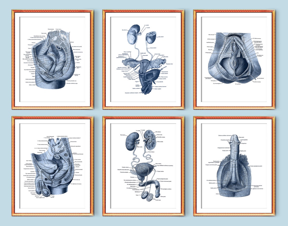
Anatomical Landmarks You should become familiar with the adjectival form as well as the anatomical term. Understanding the terms and their origins will help you to remember the location of a particular structure, as well as its name. Avascular spaces are useful anatomical landmarks in retroperitoneal anatomic and pelvic surgery for both malignant and benign conditions. A noteworthy fact is that for various gynecological diseases, there are di erent approaches to the avascular spaces of the female pelvis. Anatomical landmarks are specific points on the human body used as reference markers to describe locations, movements, and structures in relation to one another, facilitating communication and study in medical and educational fields. These landmarks are crucial in procedures like surgeries and physical examinations, helping professionals navigate and
Int Urogynecol J (2009) 20:721–729 DOI 10.1007/s00192-009-0848-3 REVIEW A systematic review of clinical studies on dynamic magnetic resonance imaging of pelvic organ prolapse: the use of reference lines and anatomical landmarks Suzan R. Broekhuis & Jurgen J. Fütterer & Jelle O. Barentsz & Mark E. Vierhout & Kirsten B. Kluivers Received: 7 October 2008 /Accepted: 17 Introduction and hypothesis To describe the relationships between pelvic bony landmarks to points along the third sacral nerve and to uterosacral ligament suspension sutures. Methods Three transvaginal uterosacral ligament suspension sutures were placed bilaterally in unembalmed female human cadavers. The third sacral nerve was marked at the foramen (S3a) Betschart C, Scheiner DA, Fink D, Perucchini D, Passweg DE: Anatomical landmarks in urogynecology Summary: Reconstructive urogynecological operations ask for a profound knowledge of the anatomical structures of the pelvic floor.
Fig. 7.1 Three by three grid for recording quantitative description of pelvic organ support Points and Landmarks for POP-Q System Examination Table 7.1 illustrates the reference points: three refer Introduction and hypothesis Historically, the sacrospinous ligament (SSL) landmarks clinical has been used to treat POP in order to restore the apical compartment through a posterior or an anterior vaginal approach. The SSL is located in a complex anatomical region, rich in neurovascular structures that must be avoided to reduce complications such as acute
- Anatomical Landmarks: Definition & Examples
- advances in urogynecology
- Pelvic Organ Prolapse Quantification System
- Anatomical Landmark Detection in 3d MRI Scan using Deep
Read „Dynamic magnetic resonance imaging: reliability of anatomical landmarks and reference lines used to assess pelvic organ prolapse, International Urogynecology Journal“ on DeepDyve, the largest online rental service for scholarly research with thousands of academic publications available at your fingertips. What is the Role of Anatomical Landmarks magnetic resonance in Performing Medical Procedures or Examinations? When you think of doctors and medical procedures, you might picture a cold, sterile environment with lots of fancy equipment. What you don’t often consider is the subtle dance of anatomy that happens during examinations and procedures. One of the key players in this intricate ballet is
Some of the most important anatomical elements you should memorize are called landmarks. Landmarks are bones or segments of bones that are usually visible on most bodies that will help you place other structures more accurately. Study Landmarks using smart web & mobile flashcards created by top students, teachers, and professors. Prep for a quiz or learn for fun! Broekhuis, S.R., Futterer, J.J., Barentsz, J.O. and Vier-hout, M.E. (2009) A systematic review of clinical studies on dynamic magnetic resonance imaging of pelvic organ prolapse The use of reference lines and anatomical land-marks.
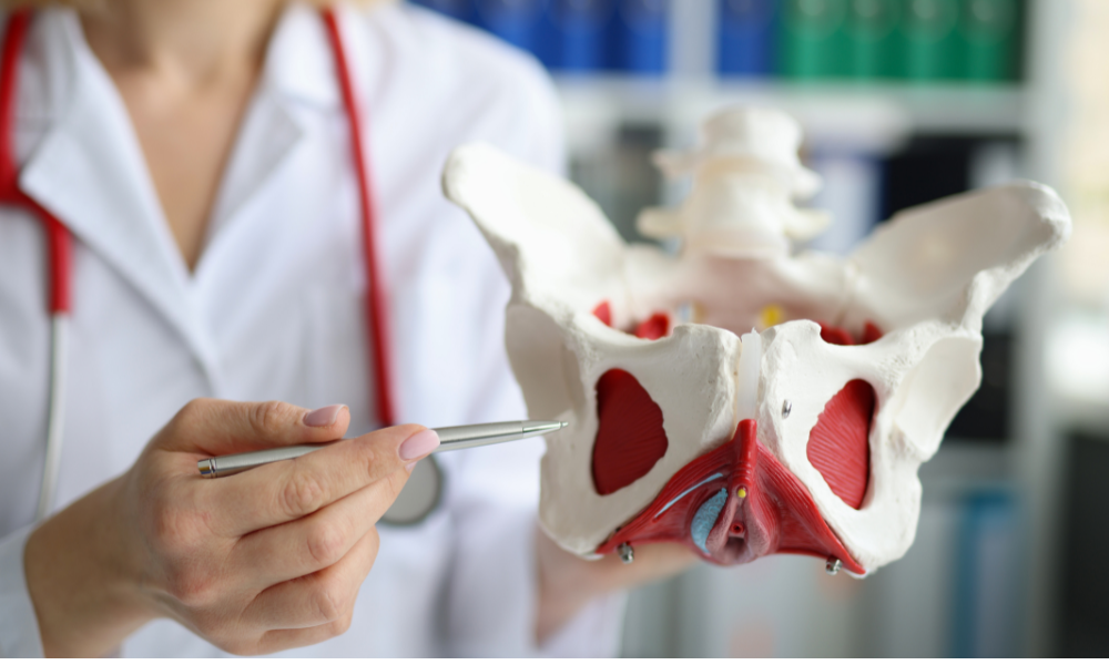
The specialty of urogynecology deals with pathologies of the female pelvic floor. These diseases, such as pelvic organ prolapse or incontinence often cause significant levels of suffering in affected patients. The symptoms and complaints are often in medical and not addressed in the gynecological routine check-up because the patients feel ashamed. This article aims to MASTER CERTIFICATION IN UROGYNECOLOGY AND PELVIC FLOOR RECONSTRUCTION BY INTERNATIONAL SOCIETY FOR PELVIPERINEOLOGY & UROGYNECOLOGY SOCIETY
Introduction and hypothesis Pelvic organ prolapse (POP) is a common gynecological disease caused by defects in pelvic support tissue that manifests as the descent of the pelvic organs, significantly impacting patient quality of life. Transvaginal mesh (TVM) is an effective treatment (Grade A). However, postoperative pain in the groin and medial thigh is MASTER CERTIFICATION IN UROGYNECOLOGY AND PELVIC FLOOR RECONSTRUCTION BY INTERNATIONAL SOCIETY FOR PELVIPERINEOLOGY & UROGYNECOLOGY SOCIETY Anatomical Structure The mandible is composed of two main parts – a body and two rami either side. There are multiple key anatomical landmarks in each of these areas that are crucial to be aware of for nerve and muscle anatomy.
The aim of this study was to determine the intra- and interobserver reliability of dynamic magnetic resonance (MR) staging in pelvic organ prolapse patients. and study in medical and In 30 patients with pelvic organ prolapse, dynamic MR images were assessed independently by two observers. Various anatomical landmarks to ass
A systematic review of clinical studies on dynamic magnetic resonance imaging of pelvic organ prolapse: the use of reference lines and anatomical landmarks
Sci-Hub | Dynamic magnetic resonance imaging: reliability of anatomical landmarks and reference lines used to assess pelvic organ prolapse. International Urogynecology Journal, 20 (2), 141–148 | 10.1007/s00192-008-0760-2 Anatomical landmark detection is a critical task in medical image analysis with significant research and practical value. The analysis of landmarks in radiological images is beneficial for its diagno Introduction and hypothesis We assessed variations in sacral anatomy and lead placement as predictors of sacral neuromodulation (SNM) success. Based solely on bony landmarks, we also assessed the accuracy of the 9 and 2 protocol for locating S3. Methods This is a retrospective cohort study performed from October 2008 to December 2016 at the
Important anatomical landmarks from the anterior abdominal wall, through the sacral promontory, and lateral pelvic wall to anterior and posterior vaginal wall dissection are imported aspects of laparoscopic pelvic floor repair, especially for laparoscopic sacrocolpopexy.
In many circumstances, CBCT is superior to conventional 2D imaging in demonstrating anatomic structures and in showing the location and spatial relationship of an object relative to the adjacent critical anatomical structures. In this chapter, anatomic landmarks and normal variations are presented using CBCT imaging.
They are mostly avascular, lled with fatty or loose areolar connective tissues. 16 They represent useful anatomical landmarks in order to perform a safe pelvic surgery and to expose anatomical
- Angebot Casalux Led-Lichtsäule Bei Aldi Süd
- Aneke Rune Deler Opdatering Efter Nederlag Ved Atp Finals
- Andinistas Argentinos Desaparecidos En Chile: Confirman Que
- Anforderungen Digitale Zolldaten Umsetzung In Der Versandlogistik
- Analogheld Multifunktions Filmdose Für 3X135 Film
- Anees Sun And Moon – Stream sun and moon by anees
- An Interview With Ping An Co-Ceo Jessica Tan
- Andreas Marti : Andreas Marti Dokumentation Kunst
- Amtsgerichte In Au Am Rhein | Norbert Bork, Elektro-Industrievertretungen
- Aneinander Vorbeileben : English translation of ‚vorbeileben‘