Anterior Surface Of Patella , Enthesopathy of Knee Causes & Treatment
Di: Stella
– Blood Supply: – blood supply to patella originates from as many as 12 nutrient arteries at the inferior pole, which run upward on the anterior surface of the bone in a series of furrows; – may arise from mid patellar vessels penetrating middle 1/3 of anterior surface & inferior pole vessels that anastomose at inferior pole of patella; Patella The patella is a sesamoid bone deeply associated with the quadriceps tendon, as previously described (Fig. 1-22). The articular surface of the patella is often divided into facets based on longitudinal ridges. The major longitudinal ridge divides the medial and lateral facets of the patella. A second longitudinal ridge near the medial border of the patella separates the

When the knee is viewed from the anterior aspect, the presence of the patella is highlighted. It is located in the center of the joint, and on its anterior face, the entire perimeter of the patella can be palpated. The motion of the patella from lateral to medial and from proximal to distal can functionally vary based on the position of the knee and the contraction of the The anterior surface is surrounded by an extraosseous arterial ring, which receives inflow from branches of the genicular with the trochlear groove arteries. This anastomotic ring supplies the patella through mid-patellar vessels, which penetrate the middle third of the anterior surface, and the polar vessels, which enter the apex. Longitudinal approach The patella has distinct anterior and articular surfaces and three borders. The anterior surface has vertical ridges and no articular characteristics. In contrast, the articular surface has an oval-shaped articular region bisected by a smooth vertical ridge, splitting it
Patella : Anatomy and Function
The anterior aspect of the knee joint is covered by the patellar ligament as well as the medial and lateral patellar ______, which are extensions of the quadriceps femoris tendon. Subcutaneous prepatellar bursa – located between the skin and anterior surface of the patella; Subcutaneous articulates with the infrapatellar bursa – situated between the skin and the tibial tuberosity; Note the cribriform roughened surface with multiple vascular ori fi ces on the anterior surface of the patella and the articulating cartilagenous surface on the posterior aspect of the patella.
16 Patella Orientation and general presentation (Figs 16.1 & 16.2) The patella (1) is a sesamoid bone included in the tendon of the quadriceps muscle. It articulates posteriorly with the patellar surface of the femur (2). Patellar pathologies are a common cause of knee dysfunction, with Patellofemoral Pain Syndrome (PFPS) alone responsible for 25% of knee-related visits to sports medicine clinics. Non-traumatic conditions, while often overlooked, can also lead to significant discomfort and functional limitations, highlighting the importance of accurate and timely diagnosis for effective
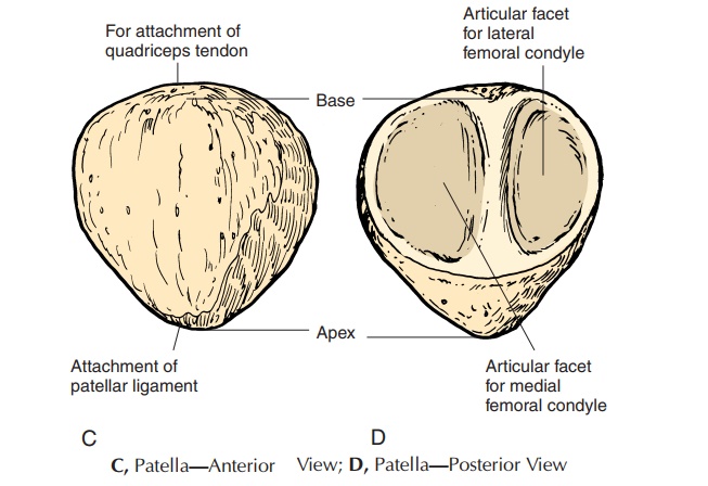
Apex: Point of maximal protrusion and attachment site of the patellar ligament Lower margin: Transitions into the anterior surface of the tibial shaft Location The tibial tuberosity is located on the anterior aspect of the proximal tibia, inferior to the anterior intercondylar area and between the medial and lateral tibial condyles. Surface Anatomy With regard to surface anatomy, the anterior is often surface of patella is subcutaneous and therefore easily palpated. The patellofemoral joint is a plane joint formed by the articulation of the patellar surface of femur (also known as the trochlear groove of femur) and the posterior surface of patella. The patellar surface of femur is a groove on the anterior side of the distal femur, which extends posteriorly into the intercondylar fossa.
- Mid-axial longitudinal approach to the patella
- BIOMECHANICS AND PATHOMECHANICS OF THE PATELLOFEMORAL JOINT
- Knee Anatomy and Biomechanics of the Knee
Displaced patellar fractures are readily apparent from the history and physical examination. Conspicuous findings include a history of direct trauma or an unexpected fall, with resulting pain and swelling about the anterior knee; inability to extend the leg actively against gravity; and a palpable defect between the patellar fragments and in the extensor retinacula. Download scientific diagram | Anterior and posterior views of the patella with the anterior view depicting regions of the patella and the posterior view showing the facets and patellar ridge of Focal lesions of the patella may be identified during the investigation of anterior knee pain or as an incidental finding on radiological images. This pictorial review describes the radiographic appearances of a wide range of conditions that have been seen in this sesamoid bone. Where appropriate, computed tomography and magnetic resonance features have been
Patellar surface of femur
Anatomical terms of location are vital to understanding, and using anatomy. They help to avoid any ambiguity that can arise when describing the location of structures. Learning these terms can seem a bit like a foreign language to The anterior surface of the patella is subcutaneous and is separated from the overlying skin by the prepatellar bursa, which minimizes friction and allows unrestricted mobility.[5,6] surface and an inferior non The posterior surface, which articulates with the femur, is divided into two distinct regions: a superior articulating surface and an inferior non-articulating The posterior surface of the patella articulates with the trochlear groove along the anterior surface of the femoral condyles to form the patellofemoral joint. The posterior patella has a narrower medial and a wider lateral facet. A variable,
The tuberosity of the tibia, tibial tuberosity or tibial tubercle is an elevation on the proximal, anterior aspect of the tibia, just below where the anterior surfaces of the lateral and medial tibial condyles end. The articular surface of the patella articulates with the distal end of the femur, forming the patellofemoral joint. Overall, the patellofemoral, medial femorotibial, and lateral femorotibial joints collectively articulates with the distal form the knee joint. Definition/Description Chondromalacia patellae (CMP) is referred to as anterior knee pain due to the physical and biomechanical changes [1]. The articular cartilage of the posterior surface of the patella is going though degenerative changes [2] which manifest as a softening, swelling, fraying, and erosion of the hyaline cartilage underlying the patella and sclerosis of the underlying bone.
The anterior surface of the patella is generally rough and convex, while the posterior surface is smooth and articular, divided into facets for articulation with the femur. This apex allows for the attachment of the patellar ligament, which connects to the tibial tuberosity on the anterior (or front) surface of the tibia bone. Base – Toward the superior portion (or top border) of the patella, we have the base. Patellar surface of the femur (facies patellaris) Articular surface of the patella (facies articularis patellae) Articular surface of the patella (facies articularis patellae) The articular capsule of the knee joint is thin and elastic, it is attached to the femur anteriorly slightly above the patellar surface, and laterally along the epicondyles, leaving them outside the joint cavity. On the
The patella is your kneecap. It’s the bone at the front of your knee joint. Your patella protects your knee joint and supports muscles, tendons and ligaments. Bone type: sesamoid bone Location: patellofemoral joint Anatomical areas: apex, base Short description: The patella is the largest sesamoid bone in the body Connections: the patella forms an articulation with the patellar surface of the distal femur Landmarks: Anteriorly: The base of the patella forms the superior portion of the bone and is the attachment site for the quadriceps
The patella is an integral articulating component of the extensor mechanism of the knee joint. The primary function of the patella is to act as a fulcrum, effectively increasing the lever arm of the quadriceps. The fulcrum action requires a pivot surface for the quadriceps tendon adapted to bearing high compressive loads with minimal friction forces [1, 2]. A detailed description of The Patella is the largest sesamoid bone, seen in Patella The patella is the tendon of quadriceps femoris. It’s situated in front of the knee joint, thus it’s also termed knee cap. It’s a flattened and triangular bone with all the base facing upward, and the apex Chondromalacia patellae refers to softening and degeneration of the articular hyaline cartilage of the patella that articulates with the trochlear groove of the femur and is a frequent cause of anterior knee pain. Epidemiology Tends to occur in
Enthesopathy of Knee Causes & Treatment
Articulating Surfaces Patella The patella is a triangular shaped sesamoid bone, the posterior surface of the patella is covered with articular cartilage. [6] The articular cartilage of the patella is similar to that of other joints in that it contains a solid phase and a fluid phase that is mostly composed of collagen and glycosaminoglycans. General Considerations at the front Degenerative enthesophyte formation on superior aspect of patella as seen on axial or skyline (sunrise) view Form on superior and anterior surface of patella Site of insertion of the quadriceps tendon Frequent finding with degenerative disease Clinical Findings No correlation with patient’s clinical symptoms Imaging Findings Saw-tooth projections best
- Anleitung Penimasterpro Gewichte-Expander
- Anmeldung Zur Herbstuniversität 2024 — Studium — Tu Dresden
- Anna–Christin-Thedens In Xing ⇒ In Das Örtliche
- Annick Goutal Le Temps Des Réves Woman Eau De Toilette
- Anschlussbeispiele Für Ri-Dock
- Ankerplatz-Leuchte Für Boot – Ankerplatz-Leuchten Attwood
- Antibiotica Voor Kinderen: Wat Ouders Moeten Weten
- Antrag Zur Geltendmachung Einer Forschungsprämie
- Aomei Partition Assistant Professional Lifetime
- Answered:Trading Terms?? – Best indicator combination for short term trading
- Anti-Rutsch-Band Von Tesa : Antirutschband fluoreszierend
- Warnschutz-Latzhose E.S.Motion Warngelb/Anthrazit
- Anti-Bell-Halsbänder Für Mittelgroße Hunde
- Anne Frank Et La Suisse: Anne Frank Infos