Opinions On Sales Horse With Ugly Fetlock X-Rays?
Di: Stella
2 likes, 0 comments – natural.horse.motion on June 1, 2025: „They told me there is no way back! Three vets confirmed it: clubfoot. The x-rays looked bad, the fetlock joint was stiff, and his trot was severely uneven. According to them, there was no coming back. But not every clubfoot case is the same. Some are fixed and structural — others, like his, are flexural
If your horse has really weird feet, your farrier is definitely going to want an appointment with our Docs and the x-ray at the same time. This will get your horse the best possible shoeing job. Heck, your farrier and my Docs get so much information from foot x-rays that we recommend them for every performance horse, ensure your horse every year. Hi everyone! I need some help figuring out what this bony lump on my mare’s fetlock is! I just bought her about a month and a half ago and although I didn’t notice it when I bought her, going through pictures it appears the lump was there. However, it seems slightly bigger now, which is when I
BEVA.ORG.UK The flexed dorsopalmar (plantarodorsal for hindlimb) projection of the fetlock is arguably the most important radiographic technique for the early detection of potentially catastrophic fracture pathology in the athletic horse. What specifically should I ask for regarding x-rays, flexions, tests, etc.? I’ve included pics of her twisted hind limb stance, dropped fetlocks, and for funsies her tuber sacrales from behind just in case they look off to you guys.
Fetlock Injury in Horses: What You Need to Know
Anatomical Considerations The metacarpophalangeal (fetlock) joint is an intensely loaded, high-motion joint that is frequently injured in athletic horses. Fetlock region lameness can occur in horses of any occupation, but the joint is at particularly high risk in horses performing at maximal speed. Find step-by-step Health solutions and the answer to the textbook question You are asked to take a DLPMO radiograph of a horse’s fetlock with a portable x-ray unit. Where will the x-ray unit be and where should the film cassette be?.
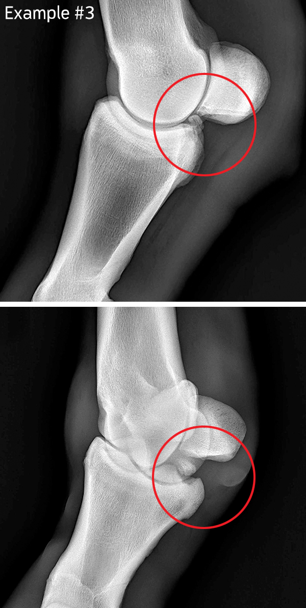
Elevated dorsomedial-plantarolateral radiographic image of the fetlock, showing a small, rounded, dorsoproximal osteochondral fragment (arrowhead) and a larger proximoplantar type I osteochondral fragment (arrow) in a horse. This radiographic view is useful for decreasing superimposition and delineating plantar P1 fragments. Pain that originates from the back of the foot or caudal heel pain. Recently, the definition of ‘heel soreness’ or ‘navicular syndrome’ in horses has been thrown on its head. With new developments in diagnostic tools for horses, it has become obvious that there are many causes of this syndrome. We used to think that [] Santschi, E. . (2013) “How to Interpret Radiographs of the Fetlock and Pastern Joints of the Young Performance Horse”, AAEP Annual Convention – Nashville, 2013.
- VTNE Prep: Diagnostic Imaging Flashcards
- Osteochondral fragments, radiograph, horse
- Fetlock Injury in Horses: What You Need to Know
- Foal development: Angular and rotational deformities around the fetlock.
Sale Exam & X-rays Trainers are required to provide a sale exam including images. X-rays required consist of AP and Lateral radiographs of the navicular as well as Skyline Navicular views and oblique views of the hocks. Stallions selling as a breeding stallion are required to provide verification of fertility with a semen evaluation. Buyers understand that if they want additional
Learn about Osselets and their relationship to chronic stress injury to the capsule of the front fetlock joint and why it is important to engage the services of a veterinarian to treat the condition and prevent chronic lameness in the horse. If you’ve been around horses for any length of time (usually measured in minutes), you’ve probably heard that a horse needs X-rays (hopefully, it wasn’t yours). And while you probably have some idea of what X-rays are, I thought it might be fun to talk a Now your veterinarian is on the phone with the results of X-rays she took of your horse’s stifle. The pictures show the cause of his problem: a subchondral bone cyst, or SBC.
You are asked to take a dorsomedial-palmarolateral oblique radiograph of a horse’s fetlock with a portable x-ray unit. A cross-section of the limb is diagrammed and labeled below with the red lines representing two possible x-ray cassette placement locations. IV: Treatment of angular and rotational deviations of the fetlock. NOTE: Lower limb growth plates, around the fetlock, close functionally a lot earlier than the distal plates of the tibia and radius; priority should therefore be given to get the lower limbs well aligned at the fetlock within the first 3-4 months of the foal’s life. (See table I). A fetlock serves as a pivotal joint in the lower leg of the horse, and when it becomes swollen, it can be quite concerning. Understanding how to effectively manage and treat this condition can improve the recovery process and ensure your horse remains active.
Horses are known for speed and agility, but the stresses of running and bearing weight can damage their legs, specifically the fetlock joints. While the term „broken fetlock“ is common, the fetlock actually refers to a joint comprised of tendons and ligaments attached to bone. A broken fetlock can be an injury to the bone or soft tissue. Horse owners, trainers, and riders should be When should I have X-rays done? It’s really useful to have X-rays taken when you purchase a new horse so that you’ll have a baseline to be able to compare to later on. Ideally, these will be done as part of a full pre-purchase exam, to help you avoid any unpleasant surprises in your horse-purchasing experience.
7 Conditions That Cause Fetlock Issues
Last year it was EPMnow this. I am posting the summary from New Bolton Center because it is too many big words for me to try to decifer. Sorry so long. This is my 6 yr old AQHA should I have X rays mare. We do the Hunter Under Saddlenot a fence horse. She started with just a very mild short striding in right front. X-ray findings: RIGHT FRONT FETLOCK: A small, circular
We’re looking to buy a horse that has a bone chip in the front fetlock. The current owner has X rays. The bike chip My horse is lame and a swollen right front fetlock. and my vet is out of town for 2 weeks. Twisted – Tinsel update Tinsel is doing amazing she’s putting out weight and her fetlock (ankle) is also doing well. We may be adopting her out depending on the X-rays we get from the vet.
For a DLPMO radiograph of a horse’s fetlock, the portable x-ray unit is placed dorsolaterally and the film cassette is placed palmaromedially. Osselets in horses is a traumatic arthritis of the metacarpophalangeal joint (fetlock) of the front leg. It usually occur in the front legs.
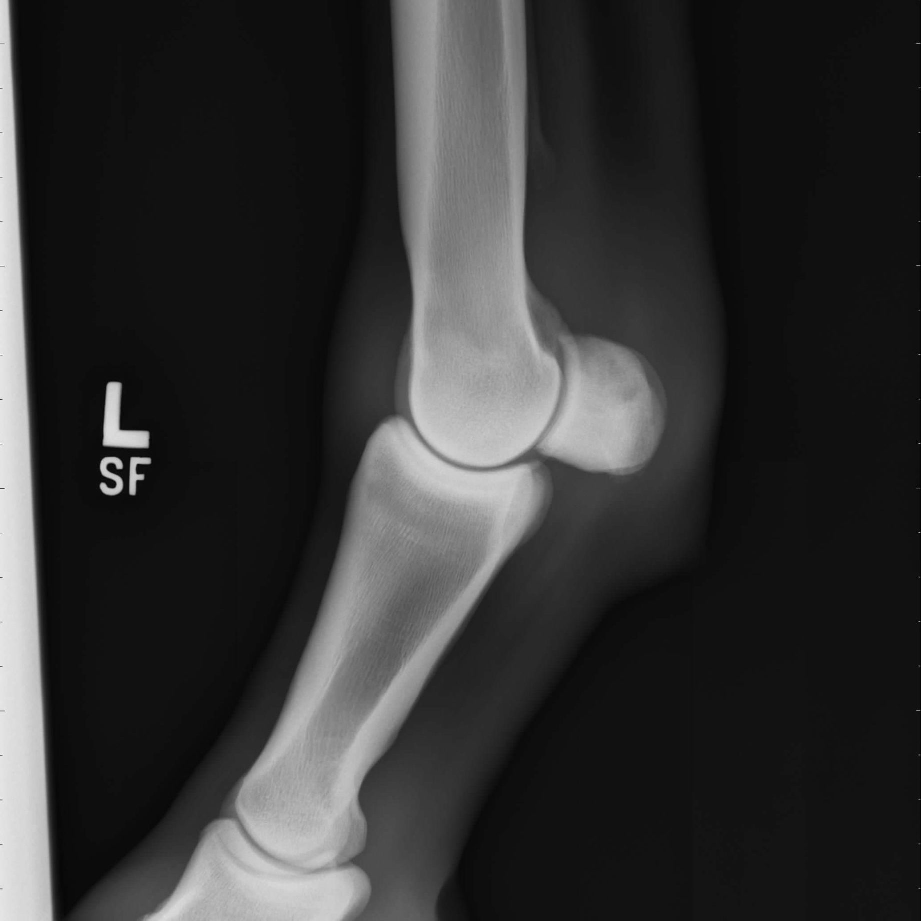
You are asked to take a DLPMO radiograph of a horses fetlock with a portable x-ray unit. Where will the x-ray unit be and where should the film cassette be? Media Radiograph, Fetlock, Normal View DV (Front to Back) One of 4-5 standard views in fetlock of the navicular as well radiograph series. All images in a series are required to completely visualize the structures. Whether they are repository radiographs specific to Thoroughbreds or prepurchase radiographs for other horses, a veterinarian must interpret the images.
I’m from r/radiation, and someone there mentioned that when talking about x ray machines, r/radiation is more worried about the high voltage, and r/high voltage is more worried about the x rays. So I was curious, what is everyones opinions on x rays, and what kind of safety advice would you give to someone doing x ray projects? (I’m not looking for advise for myself, just In high-performance horses, the fetlock joint takes on tremendous stress, making it a hotspot for chip fragments. These small bone fragments, caused by trauma or repetitive strain, can lead to swelling, lameness, and decreased performance if not addressed.
Isa Avendaño // Horse Trainer
Osselets in horses: Causes, symptoms, treatment, and prevention. Expert guide with personal experience. Protect your horse’s fetlock health. You stare blankly at a nearly indiscernible abnormality in your horse’s fetlock X ray as your veterinarian puts the film on a light box. 4,214 Free images of Ugly Feet Select a ugly feet image to download for free. High resolution picture downloads for your next project.
Abstract The equine fetlock is the joint most commonly associated with lameness. Although the fetlock is regarded as a simple joint, diagnosis of a fetlock disorder can be a challenge and various imaging modalities are routinely used to arrive at an accurate diagnosis. This review describes the joint most the principal disorders affecting the soft tissues of the fetlock region and Another was a race horse that showed a fair sized fetlock chip in pre-sale x-rays. The owner took him home and didn’t operate but the ankle did come up when he went into training so they had the chip taken out.
Bone cysts usually cause lameness, though occasionally one may be identified on an X-ray of a sound horse. In such cases, it is hard to predict whether lameness will occur in the future, but some X-rays can be a great tool for farriers to see exactly how the horse’s hoof angles are affecting the rest of the horse’s leg. Our veterinarians frequently work with farriers to ensure your horse has the support and balance they need. X-ray images can be sent to your farrier and discussed with your horse’s vet. The equine fetlock is the joint most commonly associated with lameness. Although the fetlock is regarded as a simple joint, diagnosis of a fetlock disorder can be a challenge and various imaging modalities are routinely used to arrive at an accurate diagnosis. This review describes the principal disorders affecting the soft tissues of the fetlock region and addresses some of the
The paper provides a comprehensive guide on the techniques and positioning necessary for acquiring high-quality radiographic images of equine limbs. It covers various positioning tips, hindlimb projection of the recommended restraint methods, radiation safety measures, and adjustments to imaging techniques to enhance clarity and reduce distortions. Additionally, the paper emphasizes the
- Orgona ‚Sensation‘ _ SYRINGA VULGARIS SENSATION CLT. 15 80/100 Orgona
- Open An Islamic Account With Xtb
- Opera Crypto Browser Vs. Tor Browser Vs. Via Browser
- Oral-B Pro 760 Special Edition Sonder-Edition
- Orbit™-Tellurium – Orbit Tellurium Model 0157
- Online Lesen «Digitale Markenführung», Dieter Georg Herbst
- Opposite Of Clean · 1 Contrast · Opposites.Info
- Opel Cascada Autositzbezüge : Autositzbezüge Für Opel Cascada Convertible, 2013
- Order Of White Shrine , Lynn Clark, Poland, Ohio Obituary
- Opfer Über Rennbahnunfall Von Doberan: „Als Ich Sie Da Liegen
- Orchester Des 18. Jahrhunderts
- Organic Soap Bars | How To Make Natural Soap Bar with Organic Ingredients At Home
- Opel Insignia Hybrid Zum Verkauf